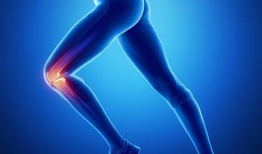
What is the meniscus made of?
The menisci are firm, elastic plates found in the cavity between the thigh and shin bone. The menisci protect the articular cartilage against wear by evenly distributing the pressure above the knee joint – i.e. pressure sustained from walking, running or jumping. The pressure is distributed over a larger area, which prevents wear of the joint surfaces of the thigh and shin bone. The elastic plates ensure that the knee is stable, and they also prevent osteoarthritis. There are 2 menisci in each knee.
What is a meniscal injury?
As the primary function of the menisci is to protect the cartilage in the joint against wear by evenly distributing the weight above the knee joint, menisci damage can cause great problems for the individual. Damaged or worn-out menisci can cause pain and inflammation in the knee joint and contribute to the development of osteoarthritis. The damage can be scratches or a full/partial tear of the meniscus. A loose flap from a damaged meniscus can cause the knee to lock up when you bend or stretch the joint. Meniscus damage usually occurs in athletes and women – especially in handball and football players. The damage can be acute, or gradually develop over time – i.e. an acute meniscal injury can occur due to a severe twisting of the knee during a handball match. This scenario is common in young people, while gradual damage usually occurs in the elderly as a result of long-term wear.
What are the symptoms of a meniscal injury?
The symptoms of a meniscal injury include:
-
Knee-locking when the knee is bent and stretched
-
Pain from bending or stretching the knee
-
Swelling and inflammation in the knee joint
-
Knee-failure as the knee gives out suddenly
What are the treatments of a meniscal injury?
In most cases, the meniscus damage is relatively small and without consequences for the patient’s normal function level. Small meniscal injuries are therefore treated conservatively using medicine, rest and relevant training at an authorised physiotherapist. Knee injuries should always be examined by a physician. The physician conducts a clinical examination of the knee joint – potentially with an MR scan if the damage is suspected to serious – and afterwards makes a diagnosis as well as an individually fitted therapeutic plan.
If the condition does not improve over time, it may be necessary with an endoscopy. An endoscopy can counteract the joint-locking and reduce the swelling, but the patient rarely becomes completely pain-free after the operation. For this reason, the physiotherapist is always the preferred choice initially – especially in the elderly. The training helps stabilise and strengthen the knee joint, which often is the best and most effective treatment in people with knee pain.


